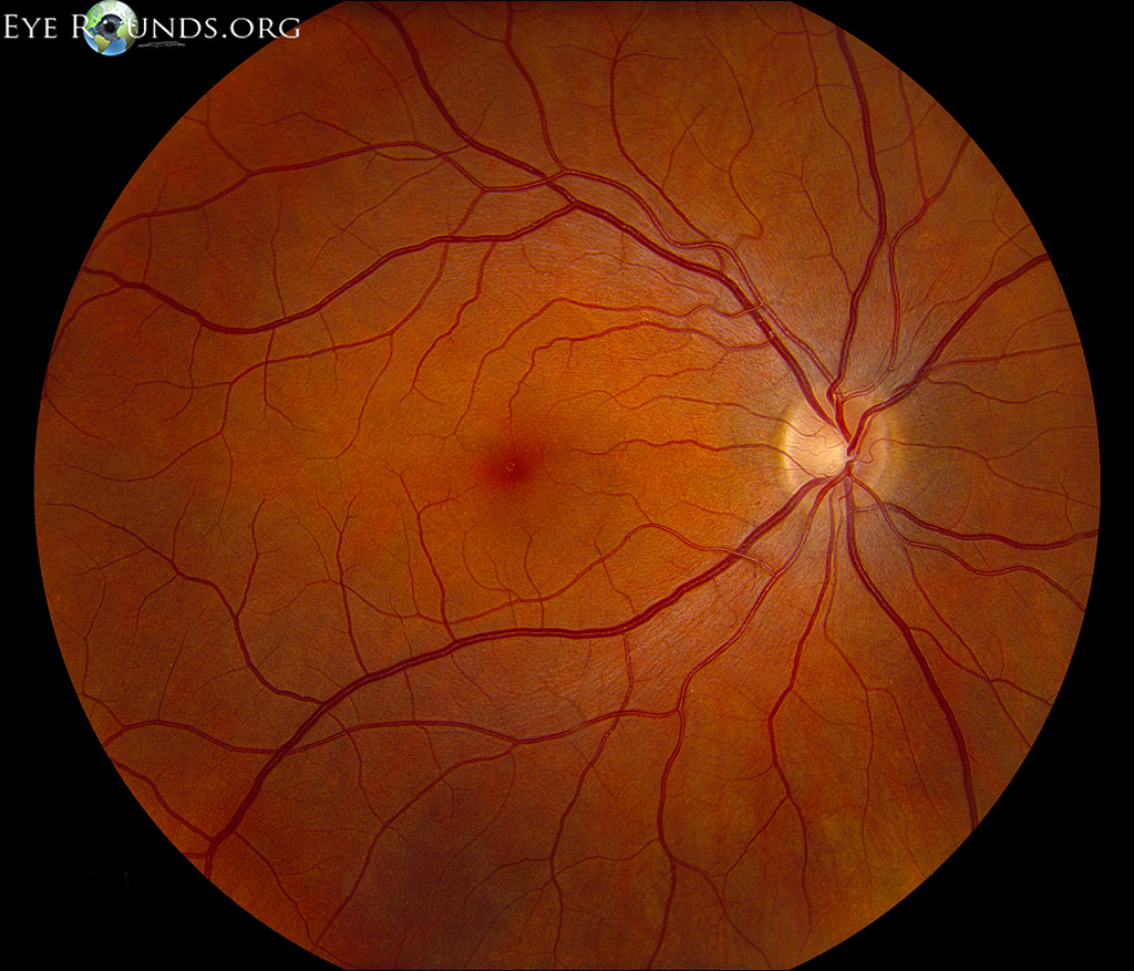
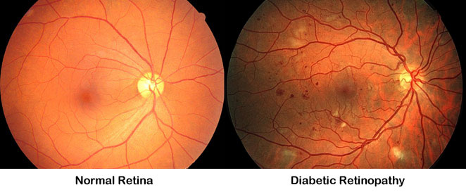
Diabetic retinopathy can cause vision loss and blindness. be proactive and stay informed. get more information on how diabetic retinopathy can progress & how it may affect you. See more videos for fundus exam diabetic retinopathy. Introduction to the fundoscopic / ophthalmoscopic exam. the retina is the only portion of the central nervous system visible from the exterior. likewise the fundus is the only location where vasculature can be visualized. so much of what we see in internal medicine is vascular related and so viewing the fundus is a great way to get a sense for.
Get Results 247
7. in patients with type i diabetes, no clinically significant retinopathy can be seen in the first 5 years after the initial diagnosis of diabetes is made. a. true. b. false. 8. in patients with type ii diabetes, the incidence of diabetic retinopathy increases with the disease duration. a. I bill medicare for fundus exam diabetic retinopathy jvn/retinopathy screening using either a diabetes dx or a hypertensive dx (since jvns will also detect hypertensive retinopathy) and then z13. 5. cpt: 92250-tc (with a 59 or xu modifier if attached to an optometry exam), 92285-tc-51. the patients do not seem to need a pre-existing diabetic retinopathy code, "just" diabetes. Features of diabetic retinopathy can include microaneurysms, intraretinal hemorrhage, exudates, cotton-wool spots, macular edema, macular ischemia, neovascularization, vitreous hemorrhage, and traction retinal detachment. symptoms may not develop until damage is advanced. test patients who have diabetic retinopathy with color fundus photography.
Ophthalmoscopy Versus Fundus Photographs For Detecting And
Patients with moderate npdr should be seen every 6 to 8 months. 2,7 there is a 12% to 27% risk that they will develop proliferative diabetic retinopathy (pdr) within 1 year. 2 the use of fundus photography is suggested for these patients, and you may obtain macular oct images at your discretion if you suspect dme. Descriptionpercentage of patients aged 18 years and older with a diagnosis of diabetic retinopathy who had a dilated macular or fundus exam performed with documented communication to the physician who manages the ongoing care of the patient with diabetes mellitus regarding the findings of the macular or fundus exam at least once within 12 months.
Source: national eye institute page updated january 2021 schedule an exam find an eyecare professional and book online in minutes! do not sell my personal information company countries. Diabetic retinopathy can occur at any age. the primary prevention and screening process for diabetic fundus exam diabetic retinopathy retinopathy varies according to the age of disease onset. several forms of retinal screening with standard fundus photography or digital imaging, with and without dilation, are under investigation as a means of detecting retinopathy. 2. grading diabetic retinopathy from stereoscopic color fundus photographs—an extension of the modified airlie house classification: etdrs report number 10. early treatment diabetic retinopathy study research roup. ophthalmology. 1991;98(5):786-806. 3. fundus photographic risk factors for progression of diabetic retinopathy: etdrs report.
The sensitivity and specificity of nonmydriatic digital stereoscopic retinal imaging in detecting diabetic retinopathy. diabetes care 2006; 29:2205. vujosevic s, benetti e, massignan f, et al. screening for diabetic retinopathy: 1 and 3 nonmydriatic 45-degree digital fundus photographs vs 7 standard early treatment diabetic retinopathy study. Diabetic retinopathy is a complication of diabetes that causes damage to the eyes. over time, the blood vessels in the back of the eye become damaged and very sensitive to light. most people notice only mild vision problems at first, but if. Diabetic retinopathy is a leading cause of blindness in american adults. due to interest in the covid-19 vaccines, we are experiencing an extremely high call volume. please understand that our phone lines must be clear for urgent medical ca. Find listings right now at topwealthinfo. com. find listings and get helpful results about your question.
The Four Stages Of Diabetic Retinopathy Modern Optometry

Learn about diabetic retinopathy (dr) early detection and a treatment option. Percentage of patients aged 18 years and older with a diagnosis of diabetic retinopathy who had a dilated macular or fundus exam performed with documented communication to the physician who manages the ongoing care of the patient with diabetes mellitus regarding the findings of the macular or fundus exam at least once within 12 months. A comparative cost analysis of digital fundus imaging and direct fundus examination for assessment of diabetic retinopathy telemed j e health. 2008 nov;14(9):912-8. doi: 10. 1089/tmj. 2008. 0013.
Diabetes Symptoms And Treatment
Abstract background: blindness due to diabetic retinopathy (dr) is the major disability in diabetic patients. although early management has shown to prevent vision loss, diabetic patients have a low rate of routine ophthalmologic examination. Diabetic retinopathy learn about the causes, symptoms, diagnosis & treatment from the merck manuals medical consumer version. please confirm that you are not located inside the russian federation the link you have selected will take you. Diabetes impacts the lives of more than 34 million americans, which adds up to more than 10% of the population. when you consider the magnitude of that number, it’s easy to understand why everyone needs to be aware of the signs of the disea. Diabetic retinopathy is a condition that affects a person with diabetes. this happens when high blood sugar levels cause damage to blood vessels in the retina. in some, the blood vessels swell and leak or can obstruct the blood flow whereas.
Check fundus exam diabetic retinopathy out our website to find what is a diabetic eye exam in your area. search for what is a diabetic eye exam. find what is a diabetic eye exam now!. Diabetic retinopathy is a complication of diabetes that causes damage to the blood vessels in the retina. learn about its causes, symptoms, and treatments here. diabetic retinopathy is blood vessel damage in the retina that happens as a res. Diabetic eye disease (diabetic retinopathy) is a complication from diabetes. symptoms of diabetic eye disease include blurry or hazy vision, difficulty focusing, and night glare from oncoming lights. learn how to prevent diabetic eye diseas.

Do you or someone you know suffer from diabetes? this is a condition in which your body doesn't produce or use adequate amounts insulin to function properly. it can be a debilitating and devastating disease, but knowledge is incredible medi. Diabetic care outlining the findings of the dilated macular or fundus exam. findings includes level of severity of retinopathy (e. g. mild nonproliferative, moderate nonproliferative, severe nonproliferative, very severe nonproliferative, proliferative) and the presence or absence of macular.
Abstract. reported here is the agreement between three examination methods chosen to detect and grade diabetic retinopathy in 124 subjects with type ii (noninsulin-dependent) diabetes mellitus. these three examination methods include ophthalmoscopy (indirect and direct) by a retina specialist, seven standard field fundus photographs read by the. Don't delay your care at mayo clinic featured conditions diabetic retinopathy is best diagnosed with a comprehensive dilated eye exam. for this exam, drops placed in your eyes widen (dilate) your pupils to allow your doctor to better view i.

Video diabetic retinopathy.
0 Response to "Fundus Exam Diabetic Retinopathy"
Posting Komentar