Examination Of The Eyes And Vision Osce Guide Youtube

Flexible Elearning Video Approach To Improve Fundus Examination
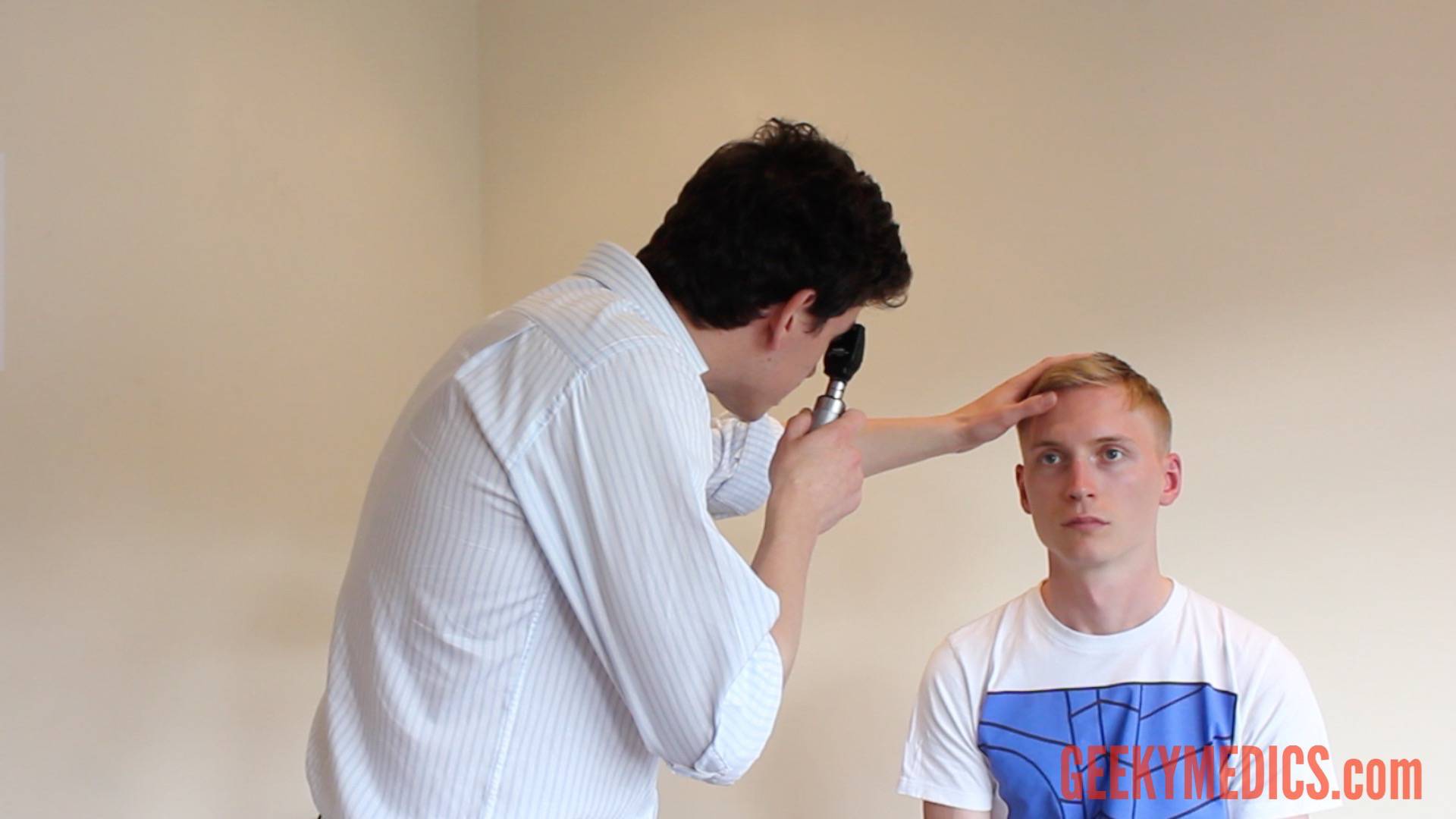
The red reflex. begin the fundoscopy examination in the patient’s “good” eye. request the patient to focus on a point in the distance. now depending on which eye you are examining first, hold the fundoscope with your right hand (to examine the patient’s right eye) or your left hand (to examine the left eye). If i could get some help to access slit lamp and 90 deg lens exam video. i greatly appreciate your attention to my request. regards, rahim. reply.
About press copyright contact us creators advertise developers terms privacy policy & safety how youtube works test new features press copyright contact us creators. Examination (video 3-1). video fundoscopy exam video 3-1 slit-lamp examination (03:17) (courtesy of richard c. allen, md, phd, facs) ophthalmoscopy. although it is difficult to accurately predict the effect of the cataract on the patient’s visual. acuity and function, the view of the fundus is usually obscured to some extent by very.
step-by-step approach to assessing the anterior eye and performing fundoscopy (ophthalmoscopy) your browser can't play this video
The d-eye video findings were compared to indirect ophthalmoscopy findings. results: the study included 172 eyes from 87 patients. in comparing d-eye to . Examination skills take repeat practice to gain proficiency and eventually perfection. the learning curve is 5-10 imaging sessions. in summary, we have provided fundoscopy exam video a walkthrough for the technique of smartphone photography and videography intended for medical students, who may not already be experts in indirect ophthalmoscopy. Click on the video icon to see a demonstration of how to hold and use the ophthalmoscope. fundoscopic abnormalities: it is important to view common .
Funduscopic Exam Video Tutorial Usmle Step 2 Cs
See more videos for fundoscopy exam. Introduction to the fundoscopic / ophthalmoscopic exam. the retina is fundoscopy exam video the only portion of the central nervous system visible from the exterior. likewise the fundus is the only location where vasculature can be visualized. so much of what we see in internal medicine is vascular related and so viewing the fundus is a great way to get a sense for.
We believe all clinicians should know how to examine the retina. this video by dr. errol ozdalga will show you how! the retina is the only portion of the cen. Aug 13, 2021 teaching medical students funduscopic examination using the flexible e-learning video approach leads to improved diagnostic accuracy of .
See our updated video here: youtu. be/yql6imge5osto see the written guide alongside the video head over to our website geekymedics. com/eye-exa. Fundoscopy (ophthalmoscopy) frequently appears in osces and you’ll be expected to pick up the relevant clinical signs using your examination skills. this guide provides a step-by-step approach to performing fundoscopy. it also includes a video demonstration. download the fundoscopy pdf checklist, or use our interactive checklist.
A step-by-step guide to performing fundoscopy (ophthalmoscopy) and assessment of the anterior eye in fundoscopy exam video an osce setting with an included video . May 26, 2014 funduscopic exam is a routine part of every doctor's examination of the eye, not just the ophthalmologist's. it consists exclusively of . Visual guide on fundoscopy by california state university.
Jove publishes peer-reviewed scientific video protocols to accelerate the first landmark observed during the funduscopic exam is the optic disc, . Apr 1, 2021 smartphone funduscopy how to use smartphone to take fundus photographs. from eyewiki. jump to:navigation, search. Compare results. find video. search for video. find it here!. This video tutorial on funduscopic exam has been provided by: doctorcallan’s channel. method of funduscopic exam: keep both yours and the person’s eyes. have the patient focus on a distant object. look at right fundus with your right eye. ophthalmoscope should be close to your eyes. your head and the scope should move together.
May 8, 2021 ophthalmoscopy (also called fundoscopy) is an exam your doctor, optometrist, or ophthalmologist uses to look into the back of your eye. This guide provides a step-by-step approach to assessing the anterior eye and performing fundoscopy (ophthalmoscopy). we've also put together a free in-depth.
0 Response to "Fundoscopy Exam Video"
Posting Komentar